Ocular Tumors
The eyes can, at times, give the first indications of tumors that are either malignant or benign and both are directly related to skin cancer — the most common being melanoma.
Melanoma is derived from a type of cell called melanocyte, which is present in the skin and is responsible for sun tanning, and, eventually sun-burning. The non-malignant form is call nevus which in lay terms is known as a "beauty spot". This represents a benign change in skin condition, composed of a clumping of melanocytes.
With melanoma, the tumor grows with little, or no, control and spreads to distant parts of the body. While the most common primary site for melanoma is the skin, it is also known to be present in other body tissues, including within the layers of the eyes. Once an ocular nevus has been identified, it must be checked on a regular basis to rule out the development of melanoma. This is because melanoma — in the early stages — often looks like a nevus, but changes with time.
The most common form of intraocular malignancy is a result of metastasis, when a cancer from a different source of the body spreads its cells to another organ. When metastasis is diagnosed — often in the form of blurred vision — it can be the first sign of cancer, especially a skin, breast or lung neoplasia.
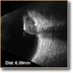
If Dr. Uniat suspects a retinal tumor, she will arrange to have the patient's pupils dilated, so that the retinas can be more effectively examined with special lenses. This can lead to more detailed examinations by a B-Scan Ultrasound or the OCT (optical coherence tomography) unit. Should these readings prove the presence of a malignant tumor, further studies with fluorescein angiography, computadorised tomography or an MRI could also be requested to evaluate the primary lesion and if spreading to other parts of the body has occurred.


