


Macular Holes
Macular holes normally occur along with aging, trauma or myopia (near-sightedness) and are usually the result of the vitreous gel in the eye pulling the tissue of the macula until it tears. As the resulting hole progresses over a period of weeks — and sometimes — months, central vision becomes blurred and distorted. Side vision, however, remains normal.
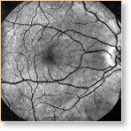
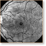
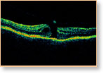
When Dr. Uniat suspects a macular hole, the patient will have their pupils dilated for detailed testing and analysis. The retinas will be visualized, firstly through special lenses, and eventually with the clinic's OCT (optical coherence tomography) unit, which provides instant readings of the structure of the eye and a clear identification of whether or not a hole is developing.
Once a hole is confirmed — and surgery is considered appropriate — an appointment will be made for Dr. Uniat to perform the operation at the Royal Alexandra Hospital. In vitrectomy surgery, Dr. Uniat uses delicate instruments inside the eye to remove the vitreous gel that is pulling on the macula. This space is then filled with a special gas bubble, which absorbs over a period of about 2-6 weeks. This gas is progressively replaced by a fluid produced by the eye.
To have any chance of optimum results of this surgery, however, the patient must remain in face-down position for up to two weeks to maintain the gas bubble's direct contact with the macula.
The extent of success will also depend, to some extent, on the amount of time between the hole becoming established and the surgical operation.