

|
||
Retina Diseases |
||
Detached Retina
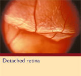
The retina is the inner layer of the eye ball, and may sometimes get split from the outer layers, and become detached. Detached retinas are more likely to occur after middle age and affect people who are near-sighted. How does the retina become detached?Tiny tears or holes in the retina are usually caused by aging of tissues in the eye. The eye largely maintains its round shape from the gel-like vitreous that fills its interior. Aging often causes the vitreous to shrink, and some of the fine fibres, linked to the retina, may be pulled free - causing a vitreous detachment. This can cause tears or holes in the retina, which allows fluid to get under the retina, leading to the detachment.
Some symptomsWhile it is normal to see "floaters" in the eye, if there is a sudden increase in the floaters or development of flashes or cobwebs, this may be an indication of a retinal tear. A much more serious condition is if the retina becomes detached. This happens when the flue gets under the retina and it starts a peeling process. As this peeling process reaches the central zone (the macula) the vision loss will be dramatic - and urgent surgery is required. TreatmentFor some new or small tears, before a detachment has started, the treatment will likely be limited to the use of a laser or cryotherapy. However, if a retinal detachment has occurred, all the holes in the retina must be sealed. Extensive detachment requires major eye surgery, usually in the Royal Alexandra Hospital. Among the procedures and appliances that might be used in treatment are:
PrognosisMore than 90% of detachments can be successfully treated, with the retina re-attached with one procedure or surgery. It is vital, however, that treatment takes place before the central part of the retina - the macula - becomes detached. Once the macula has been detached for a few days, there is less chance of a complete return of central vision. |
||

